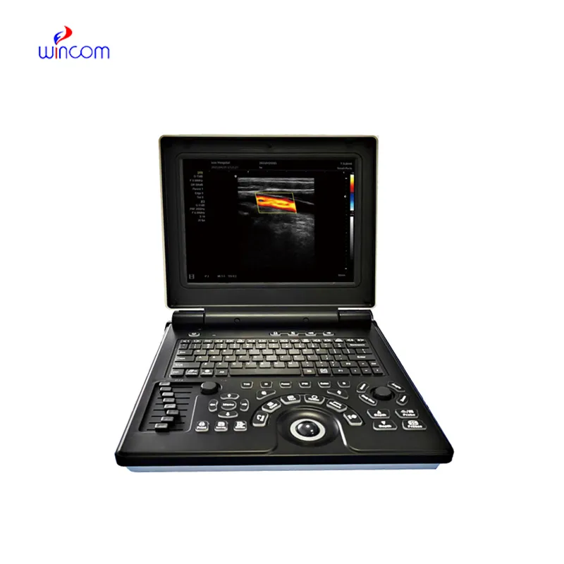
With video displayed through beamforming and noise-filtering technology, the baby fetal doppler is able to present an image that is very sharp and stable. Easy-to-use touch-screen controls help to streamline the process while rapid image rendering is guaranteed by the fast processing. Equipped for contemporary healthcare settings, the baby fetal doppler is capable of working with both 2D and Doppler imaging.

The baby fetal doppler is a very significant diagnosis tool used in obstetrics for fetal monitoring and pregnancy development detection. It indirectly affects the cardiology field by providing information about the hearts and blood flow dynamics. Furthermore, the baby fetal doppler is very important in diagnosing abdominal problems, especially issues with the liver, kidneys, and gallbladder. Still, it is also being used in musculoskeletal diagnoses for spotting ligament and tendon injuries.

The baby fetal doppler will proceed to develop as new innovations emerge in artificial intelligence and data analysis. The new models of the baby fetal doppler will be able to provide training simulations that experts can use to improve scanning sessions. The increased processing power and connectivity of the baby fetal doppler will set new standards of accessibility and accuracy in medical scanning.

For long-term functionality, it is recommended that the baby fetal doppler remain within an environment that maintains controlled levels of both humidity and temperature. The cables should be unwound slowly to ensure that no undue stress or wire breakages occur. The baby fetal doppler should also be properly disinfected each time a patient has been examined.
Designed to be accurate and functional, the baby fetal doppler offers high-definition imaging for diagnosis in numerous medical settings. It is comfortable with obstetric, vascular, and abdominal procedures and delivers exceptional definition. The baby fetal doppler increases the certainty of diagnosis and reduces patient disruption through its non-invasive mode of operation. Its digital components allow for storage of data, transfer of images, and analysis.
Q: What makes the ultrasound scannert effective for diagnostic imaging? A: Its high-frequency sound wave technology allows accurate visualization of internal body structures in real time. Q: How portable is the ultrasound scannert? A: The device features a compact and lightweight design, allowing easy movement between clinical departments. Q: What types of probes are compatible with the ultrasound scannert? A: It supports multiple probe types, including linear, convex, and phased array probes for varied diagnostic needs. Q: Does the ultrasound scannert require special training to operate? A: Basic technical training is recommended to maximize its imaging performance and functionality. Q: How long can the ultrasound scannert operate continuously? A: It is designed for extended use with efficient cooling systems and stable power performance.
The centrifuge operates quietly and efficiently. It’s compact but surprisingly powerful, making it perfect for daily lab use.
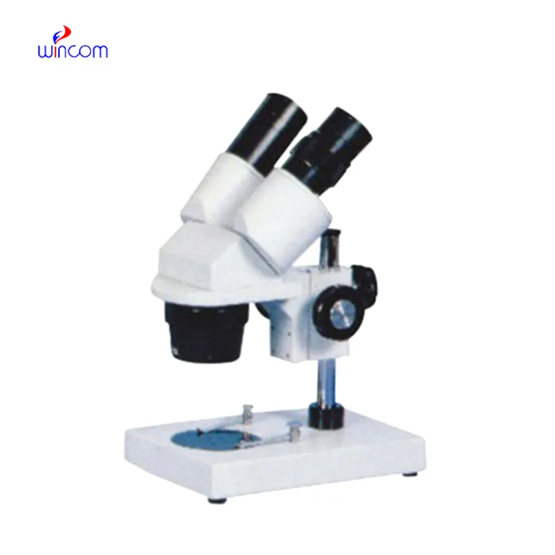
The microscope delivers incredibly sharp images and precise focusing. It’s perfect for both professional lab work and educational use.
To protect the privacy of our buyers, only public service email domains like Gmail, Yahoo, and MSN will be displayed. Additionally, only a limited portion of the inquiry content will be shown.
We are planning to upgrade our imaging department and would like more information on your mri machin...
I’m looking to purchase several microscopes for a research lab. Please let me know the price list ...
E-mail: [email protected]
Tel: +86-731-84176622
+86-731-84136655
Address: Rm.1507,Xinsancheng Plaza. No.58, Renmin Road(E),Changsha,Hunan,China

 af
af
 es
es
 ar
ar
 tr
tr
 sw
sw
 pt
pt
 th
th
 ur
ur
 bn
bn
 ne
ne
 vi
vi
 km
km
 lo
lo
 de
de
 ru
ru
 fi
fi
 nl
nl
 fa
fa
 fr
fr
 ko
ko