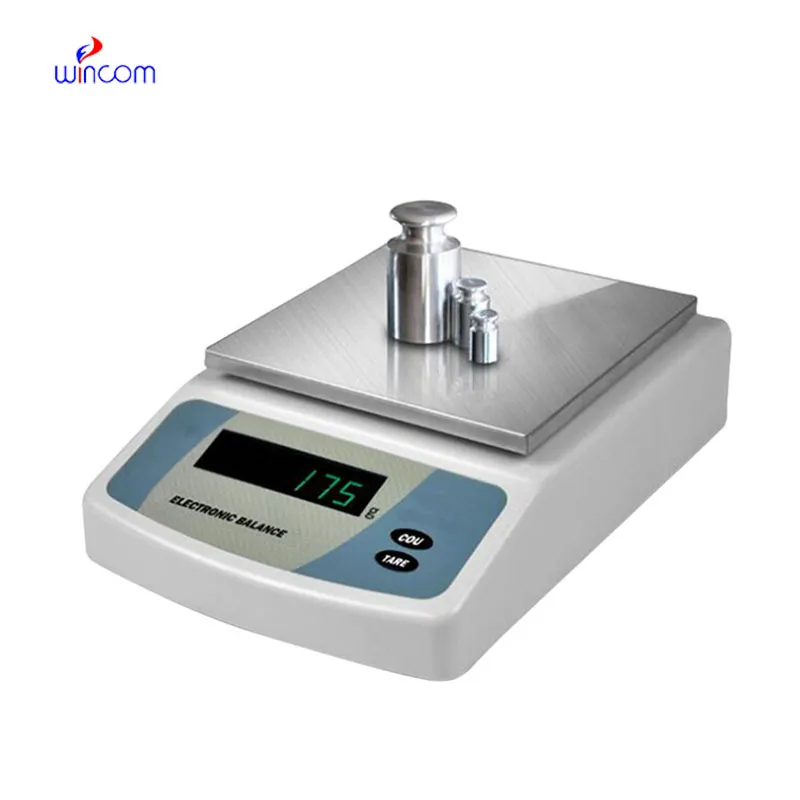
The use of balancing electrons in pharmaceutical laboratories is crucial for the accurate measurement of both active substances and excipients. Its extraordinary accuracy eliminates the possibility of formulation errors and makes regulatory compliance easier. balancing electrons are employed by laboratory staff for daily quality control, validation of batches, and research activities. Adding balancing electrons to the laboratory operations not only the consistency but also the reproducibility and the accuracy of the results for clinical trials and research applications are assured.

In research labs of the biomedical field, balancing electrons is used while standardizing the experimental samples. For the purpose of testing, h researchers have to measure the biological or chemical samples very accurately and in this way, they do not use more than the required amount of sample for analytical testing. This process keeps the studies that compare different methodologies consistent and at the same time it prevents different results that are due to the difference in the samples’ mass. By providing correct input values, balancing electrons makes it easier for the experimenters to repeat the experiments and to trust the data more in the hospitals’ research institutions.
The future application of balancing electrons will be broadened in education laboratories at teaching hospitals. The training provided to lab techs and medical researchers will be accomplished with the help of advanced simulation modes and guided measurement functions. This revolution will offer the medical student the chance to learn practically while the doctor will continue to rely on the precision of the instruments in the lab.
The maintenance of balancing electrons involves the aspects of storage and inactivity care that come first. The balance should be protected from dust and vibration when it is not in active use. Periodically checking the operational status during long storage prevents unnoticed performance drift. These practices guarantee that balancing electrons is still capable of accurate use in laboratories, medical and hospital settings.
balancing electrons is an essential teaching device in the training of laboratory and hospital technicians. It allows students and trainees to practice accurate weighing, calibrating, and sample-handling methods properly. balancing electrons is the way to take a tour through precision measurement and correct lab practices, and at the same time, to gain support in the formation of skilled manpower for the clinical, pharmaceutical, and research labs.
Q: What distinguishes an Analytical Balance from a precision balance? A: The analytical balances have a higher sensitivity and a finer readability for measuring masses of very small amounts. Q: Is an Analytical Balance appropriate for pharmaceutical applications? A: It is widely used for weighing active ingredient and formulation components. Q: Is it mandatory for an Analytical Balance to have a draft shield? A: Draft shields have the function to prevent air disturbances which might affect the weighing results. Q: What are the possible types of materials that can be weighed on an Analytical Balance? A: Weighing of powders, chemicals, and biological samples, as well as reference weights are the most common measurement. Q: Is it possible for several users to work with the same Analytical Balance? A: Yes, but the proper handling procedures and access controls must be strictly adhered to.
The hospital bed is well-designed and very practical. Patients find it comfortable, and nurses appreciate how simple it is to operate.
The delivery bed is well-designed and reliable. Our staff finds it simple to operate, and patients feel comfortable using it.
To protect the privacy of our buyers, only public service email domains like Gmail, Yahoo, and MSN will be displayed. Additionally, only a limited portion of the inquiry content will be shown.
We are planning to upgrade our imaging department and would like more information on your mri machin...
We’re currently sourcing an ultrasound scanner for hospital use. Please send product specification...
E-mail: [email protected]
Tel: +86-731-84176622
+86-731-84136655
Address: Rm.1507,Xinsancheng Plaza. No.58, Renmin Road(E),Changsha,Hunan,China

 af
af
 es
es
 ar
ar
 tr
tr
 sw
sw
 pt
pt
 th
th
 ur
ur
 bn
bn
 ne
ne
 vi
vi
 km
km
 lo
lo
 de
de
 ru
ru
 fi
fi
 nl
nl
 fa
fa
 fr
fr
 ko
ko