The handheld dental x ray machine has been designed keeping in mind the needs of modern healthcare. The system comes equipped with capabilities such as real-time imaging adjustments and intelligent exposure. The digital detector provides high uniformity of images, thus supporting accurate interpretations. The handheld dental x ray machine comes with connectivity solutions that ensure smooth integration with digital radiology networks.

The handheld dental x ray machine is commonly used in medical imaging to examine skeletal trauma, lung disease, and dental anatomy. The handheld dental x ray machine assists physicians in diagnosis of fractures, infection, and degenerative disease. The handheld dental x ray machine is also used in orthopedic surgery intraoperatively. In emergency medicine, it provides rapid diagnostic information that allows clinicians to assess trauma and internal injury rapidly.

Future versions of the handheld dental x ray machine will combine energy-efficient technology with high-resolution imaging. Predictive analytics integration will enable early disease detection and personalized screening. Global telemedicine networks will also be enabled by the handheld dental x ray machine, extending access to diagnosis in underserved populations.
The life of the handheld dental x ray machine relies on proper maintenance and surveillance. The X-ray tube, generator, and control panel are some of the parts that need to be examined and serviced based on manufacturer recommendations. The handheld dental x ray machine should be protected from moisture, vibration, and heavy dust to prevent performance loss.
The handheld dental x ray machine is an important part of the healthcare system as it provides real-time imaging services for internal exams. The handheld dental x ray machine provides high-quality images that help in detecting structural anomalies. The handheld dental x ray machine is used extensively in hospitals and research institutes for bone density scans, lung scans, and dental scans.
Q: What makes an x-ray machine different from a CT scanner? A: An x-ray machine captures a single 2D image, while a CT scanner takes multiple x-rays from different angles to create 3D cross-sectional views. Q: How is image quality measured in an x-ray machine? A: Image quality depends on factors like contrast, resolution, and exposure settings, which are adjusted based on the target area being examined. Q: What power supply does an x-ray machine require? A: Most x-ray machines operate on high-voltage power systems, typically between 40 to 150 kilovolts, depending on their intended use. Q: Can x-ray machines be used for dental imaging? A: Yes, specialized dental x-ray machines provide detailed images of teeth, jaws, and surrounding structures to support oral health assessments. Q: How does digital imaging improve x-ray efficiency? A: Digital systems allow instant image preview, faster diagnosis, and reduced need for retakes, improving workflow efficiency in clinical environments.
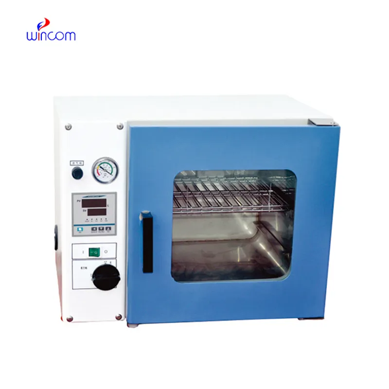
The centrifuge operates quietly and efficiently. It’s compact but surprisingly powerful, making it perfect for daily lab use.
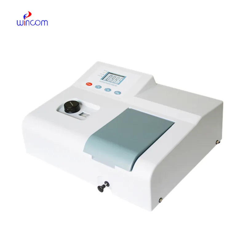
The microscope delivers incredibly sharp images and precise focusing. It’s perfect for both professional lab work and educational use.
To protect the privacy of our buyers, only public service email domains like Gmail, Yahoo, and MSN will be displayed. Additionally, only a limited portion of the inquiry content will be shown.
Could you share the specifications and price for your hospital bed models? We’re looking for adjus...
Could you please provide more information about your microscope range? I’d like to know the magnif...
E-mail: [email protected]
Tel: +86-731-84176622
+86-731-84136655
Address: Rm.1507,Xinsancheng Plaza. No.58, Renmin Road(E),Changsha,Hunan,China

 af
af
 es
es
 ar
ar
 tr
tr
 sw
sw
 pt
pt
 th
th
 ur
ur
 bn
bn
 ne
ne
 vi
vi
 km
km
 lo
lo
 de
de
 ru
ru
 fi
fi
 nl
nl
 fa
fa
 fr
fr
 ko
ko