With the cutting-edge imaging processors, the handheld fetal doppler facilitates real-time, high-resolution images that are paramount in the detection of subtle physiological changes by clinicians. The display is user-friendly for easy parameter modification as well as image marking. The handheld fetal doppler exhibits the mix of effectiveness, mobility, and reliability for a huge range of diagnostic procedures.

The handheld fetal doppler has a very wide use in radiology where it supports the guidance of non-invasive procedures. It is very important in gynecology where it is allowed to conduct reproductive system evaluations. In orthopedics, the handheld fetal doppler helps visualize muscles, joints, and tendons ensuring correct diagnostic interpretation. It is this very nature of versatility that makes it a must-have for imaging during procedures in real time.

The handheld fetal doppler can look forward to getting advantages from miniaturization and wearable technologies. Portable or handheld versions of the handheld fetal doppler will become more widespread to facilitate quick diagnoses in rural as well as emergency setups. The integration of telemedicine services will thus facilitate concurrent consultations via the handheld fetal doppler.

Proper care and management of the handheld fetal doppler needs to be carried out to ensure that it functions well at all times. Cleaning of the probes using a recommended disinfectant helps prevent contamination of the probe and image distortion. Storage of the handheld fetal doppler in a clean and dry place away from high temperatures helps prolong the life of the equipment.
With advanced transducer technology, the handheld fetal doppler delivers reliable and high-resolution imaging capability. It allows healthcare providers to see anatomical structures with unmatched clarity and speed. The handheld fetal doppler enhances workflow efficiency in hospitals, clinics, and mobile medical facilities. With rugged construction and digital connectivity, it makes integration into existing healthcare systems easy.
Q: What are the main maintenance requirements for the ultrasound scannert? A: Regular cleaning, proper probe handling, and scheduled inspections help maintain optimal performance. Q: How often should the ultrasound scannert be calibrated? A: Calibration frequency depends on usage levels, but periodic professional checks are recommended. Q: Is the ultrasound scannert suitable for pediatric use? A: Yes, it provides gentle, non-invasive imaging ideal for neonatal and pediatric diagnostics. Q: Does the ultrasound scannert support wireless connectivity? A: Many models include Wi-Fi or Bluetooth features for data sharing and device integration. Q: What materials are used in the ultrasound scannert construction? A: It is built with durable medical-grade components designed to withstand continuous clinical use.
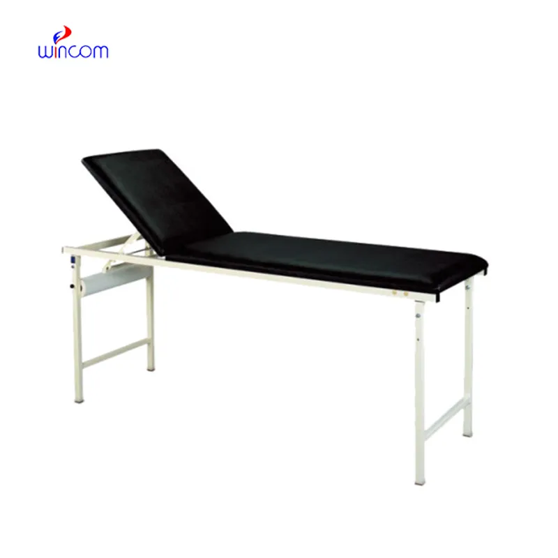
The hospital bed is well-designed and very practical. Patients find it comfortable, and nurses appreciate how simple it is to operate.
We’ve used this centrifuge for several months now, and it has performed consistently well. The speed control and balance are excellent.
To protect the privacy of our buyers, only public service email domains like Gmail, Yahoo, and MSN will be displayed. Additionally, only a limited portion of the inquiry content will be shown.
Hello, I’m interested in your water bath for laboratory applications. Can you confirm the temperat...
I’d like to inquire about your x-ray machine models. Could you provide the technical datasheet, wa...
E-mail: [email protected]
Tel: +86-731-84176622
+86-731-84136655
Address: Rm.1507,Xinsancheng Plaza. No.58, Renmin Road(E),Changsha,Hunan,China

 af
af
 es
es
 ar
ar
 tr
tr
 sw
sw
 pt
pt
 th
th
 ur
ur
 bn
bn
 ne
ne
 vi
vi
 km
km
 lo
lo
 de
de
 ru
ru
 fi
fi
 nl
nl
 fa
fa
 fr
fr
 ko
ko