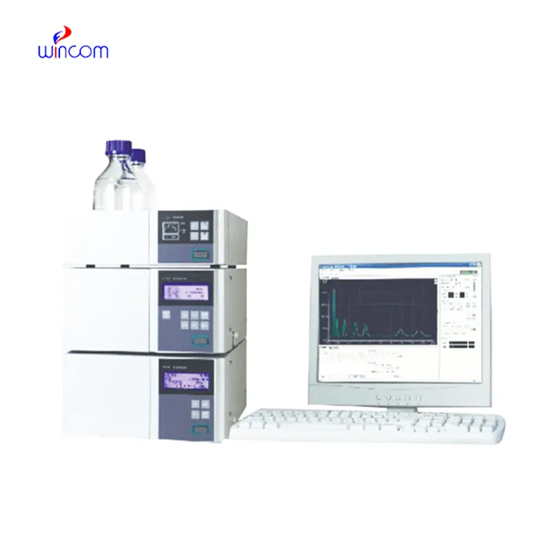
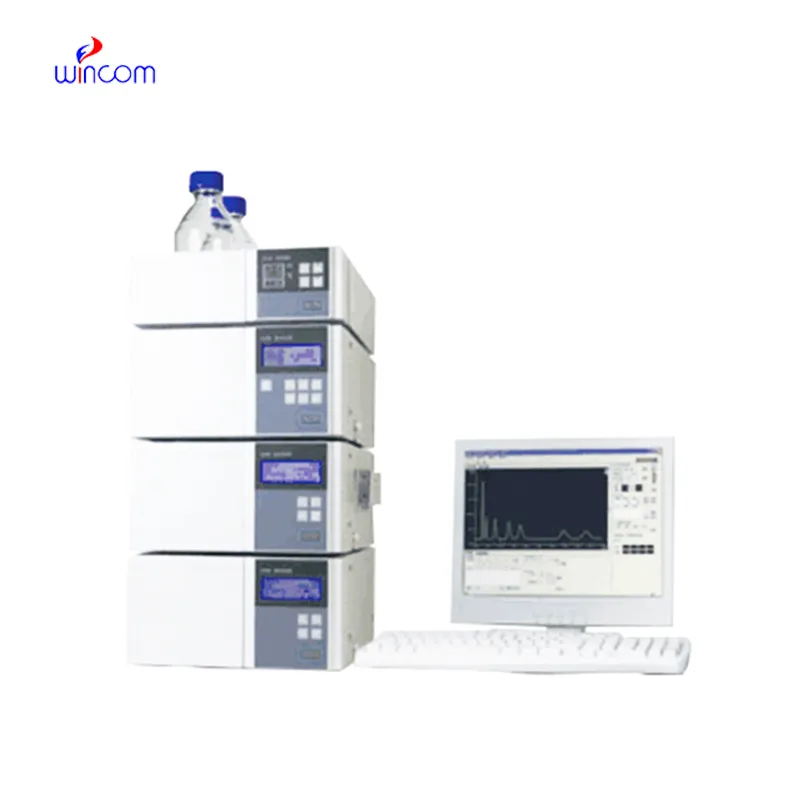
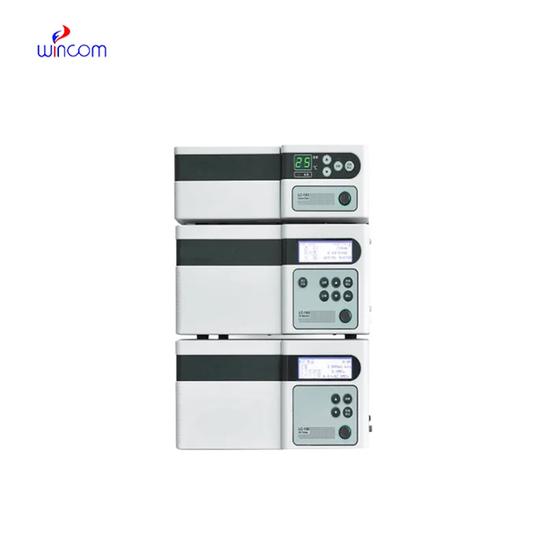
In the pharmaceutical lab, prep hplc is the key to the precise assessment of the active substances, impurities, and metabolites. The machine gives a high-resolution separation, which in turn supports the quality assurance and the regulation compliance. Lab workers put their trust on prep hplc for method validation, production consistency monitoring, and research trials. prep hplc brings together the delicate ability to detect plus the repeated nature of results to make the complex formulations proficiently analyzed, thus, it serves the routine lab testing and the advanced experimental work in hospitals, research centers, and clinical facilities both.
The quality control process for prep hplc in intravenous medications and hospital-prepared solutions is being carried out by hospital laboratories. It isolates the impurities and analyzes the active substances to ascertain the uniformity of the composition. This practice enables the pharmacists and laboratory staff to verify the drug's quality before it gets to the patient, hence minimizing the risk associated with it and at the same time endorsing the safe therapeutic practices in hospitals.
In hospitals and clinical research, prep hplc techniques will get higher resolution columns and ultrafast chromatography methods more and more. It will be possible to do these innovations in a shorter time and with a more accurate result. Future prep hplc applications will be used to identify biomarkers quickly, monitor therapies in real-time, and manage patients more efficiently in both the laboratory and clinical settings.
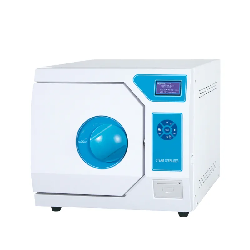
prep hplc will require regular maintenance to be kept up in order to continue providing precise measurements in medical laboratories. After every use, the technicians should flush the columns, check the seals, and inspect the tubing for wear and tear and ensure that the detector is working. Regular calibration and good solvent management decrease the chances of system damage and increase the consistency of the results. Good care and maintenance not only increase the efficiency of the laboratory but also help in providing reliable diagnostics and maintaining the instruments for hospital applications.
prep hplc is of utmost importance in biochemistry laboratories of both universities and hospitals. It makes detailed study of proteins, peptides, and metabolites possible through the separation of intricate mixtures. The application of it includes but is not limited to enzymatic analysis, biomarker detection, and data obtained through metabolomics. The sensitivity and reproducibility of the device guarantee genuine molecular profiles. Lab technicians make use of prep hplc to conclude their experiments and provide evidence for scientific publications. Its accuracy and versatility give biochemistry labs the ability to perform cutting-edge research in molecular mechanisms, disease pathways, and therapy targets thus, it becomes an indispensable tool for both analytical and clinical lab investigations.
Q: What is HPLC used for in laboratories? A: HPLC turns out to be one of the most significant and essential analytical methods in laboratories equipped with the chemical compound analysis, separation, identification, and quantification of their presence in complex samples which are the research, clinical, and pharmaceutical applications. Q: How does HPLC separate compounds? A: The HPLC separation technique is based on the different affinities of the compounds to the stationary phase and mobile phase within the chromatography column. Q: Can HPLC analyze biological samples? A: Yes, it is certainly possible to carry out analyses on various biological fluids such as blood, serum, urine, etc. for the detection of metabolites, drugs, and biomarkers. Q: How often should HPLC columns be replaced? A: The replacement of the columns must be done according to the manufacturer instructions or when the performance begins to decline, which is quite usual after heavy use or contamination. Q: What detectors can be used with HPLC? A: The analysis type determines the use of, among others, UV, fluorescence, refractive index, and mass spectrometry detectors as the common detectors.
The centrifuge operates quietly and efficiently. It’s compact but surprisingly powerful, making it perfect for daily lab use.
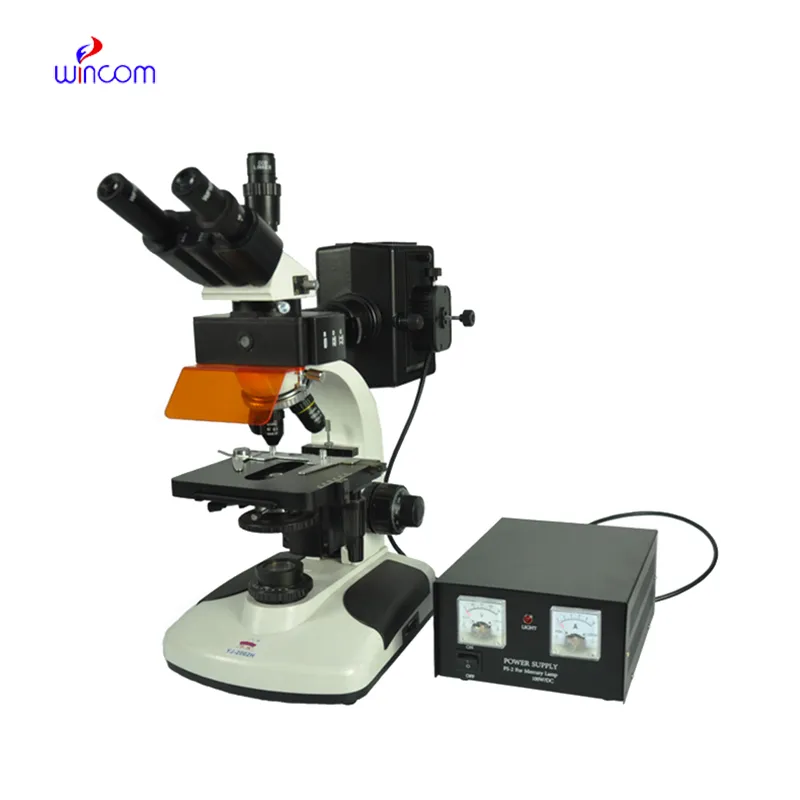
I’ve used several microscopes before, but this one stands out for its sturdy design and smooth magnification control.
To protect the privacy of our buyers, only public service email domains like Gmail, Yahoo, and MSN will be displayed. Additionally, only a limited portion of the inquiry content will be shown.
I’d like to inquire about your x-ray machine models. Could you provide the technical datasheet, wa...
I’m looking to purchase several microscopes for a research lab. Please let me know the price list ...
E-mail: [email protected]
Tel: +86-731-84176622
+86-731-84136655
Address: Rm.1507,Xinsancheng Plaza. No.58, Renmin Road(E),Changsha,Hunan,China

 af
af
 es
es
 ar
ar
 tr
tr
 sw
sw
 pt
pt
 th
th
 ur
ur
 bn
bn
 ne
ne
 vi
vi
 km
km
 lo
lo
 de
de
 ru
ru
 fi
fi
 nl
nl
 fa
fa
 fr
fr
 ko
ko