stationary phase in hplc hangs the hospital laboratory in the sense of getting quick and reproducible results for patient sample analysis. Its use is widespread to separate small molecules, hormones, and therapeutic drugs with pinpoint accuracy. Lab staff apply stationary phase in hplc in discovering biomarkers, doing pharmacokinetic studies, and metabolite profiling. Its flexibility makes it suitable for clinical applications with different requirements like research, routine diagnostics, and patient care. So, when hospitals include stationary phase in hplc into their laboratory processes, they get not only the speed but also the dependable analytical performance over various departments.
The hospital laboratory technicians employ stationary phase in hplc to quantify the quantity of proteins or peptides. This assists in the research of biomarkers, immunotherapy studies, and analysis of responses induced by novel therapies among patients. Its accuracy and sensitivity enable the obtaining of correct results, hence aiding superior research.
In hospitals and clinical research, stationary phase in hplc techniques will get higher resolution columns and ultrafast chromatography methods more and more. It will be possible to do these innovations in a shorter time and with a more accurate result. Future stationary phase in hplc applications will be used to identify biomarkers quickly, monitor therapies in real-time, and manage patients more efficiently in both the laboratory and clinical settings.
Regular system checks, cleaning of detector flow cells, and changing consumable parts whenever necessary are some of the actions that the laboratory staff should take in order to keep the stationary phase in hplc working efficiently. Observing pump performance, taking care of solvent contamination, and storing columns correctly prolong the life of the instrument. Good maintenance assures reproducibility, cuts down on time without access to equipment, and promotes high-quality analysis in hospitals and clinical labs.
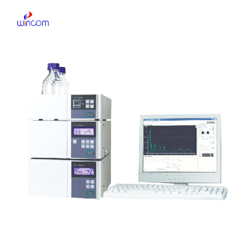
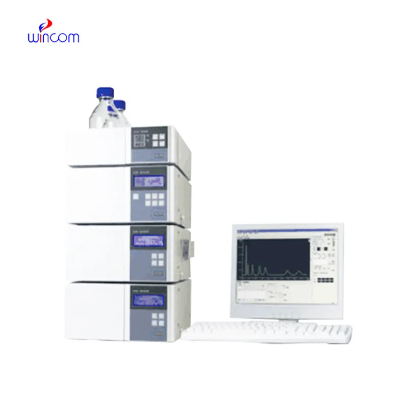
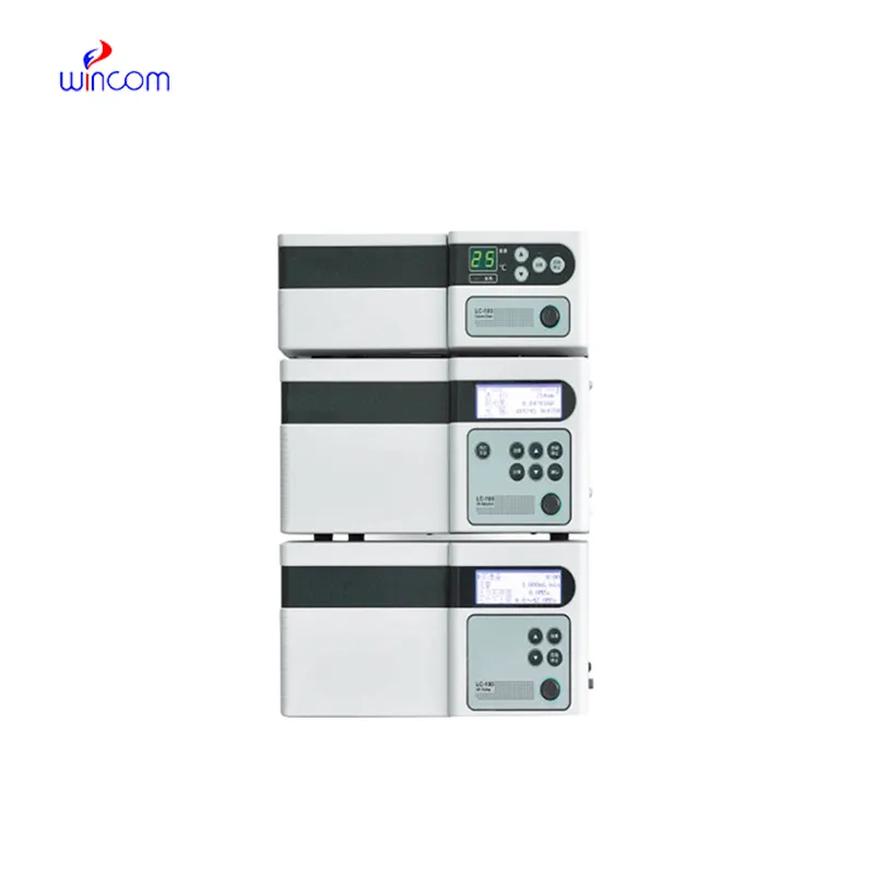
In today's laboratories, stationary phase in hplc is indispensable for chemical analysis and serves as a primary instrument. Detection of compounds in intricate mixtures is first done through separation and then identification. Consequently, researchers can precisely check the interactions between molecules. stationary phase in hplc is regarded to have extremely high reproducibility and it shares its strength with the fields of pharmaceuticals, biochemistry, and environmental science. Its alliance with sensitive detectors leads to the accurate measurement of very small amounts. stationary phase in hplc is the trustworthy partner of lab technicians in validation of experiments, profiling of samples, and development of analytical methods. It not only gives consistent and detailed results but also boosts the efficiency of laboratories and at the same time, makes sure that the data obtained from research is reliable and thus, supports the advanced scientific inquiries that are conducted in various disciplines.
Q: What is the sample preparation for HPLC? A: For the most part, samples should be filtered, diluted, or subjected to solvent extraction in order to avoid column clogs and have the results be accurate Q: Is HPLC able to pick trace-level compounds? A: With the right detectors, it can pick up such substances in extremely small amounts with high sensitivity. Q: Is HPLC a method that can be applied to analysis of proteins? A: Yes, particularly if one employs size-exclusion and reversed-phase columns for protein, peptide, and biomolecule separation. Q: What is the process of calibrating HPLC? A: The process is done by taking standards of known concentrations that are the same as the one in the sample and using them to check the performance of the column and the accuracy of the detector. Q: Are particular solvents needed for HPLC? A: Yes, the solvents used need to be compatible with the type of the column and the detectors to prevent any damage or interference in the analysis process.
The hospital bed is well-designed and very practical. Patients find it comfortable, and nurses appreciate how simple it is to operate.
The centrifuge operates quietly and efficiently. It’s compact but surprisingly powerful, making it perfect for daily lab use.
To protect the privacy of our buyers, only public service email domains like Gmail, Yahoo, and MSN will be displayed. Additionally, only a limited portion of the inquiry content will be shown.
Could you share the specifications and price for your hospital bed models? We’re looking for adjus...
Could you please provide more information about your microscope range? I’d like to know the magnif...
E-mail: [email protected]
Tel: +86-731-84176622
+86-731-84136655
Address: Rm.1507,Xinsancheng Plaza. No.58, Renmin Road(E),Changsha,Hunan,China

 af
af
 es
es
 ar
ar
 tr
tr
 sw
sw
 pt
pt
 th
th
 ur
ur
 bn
bn
 ne
ne
 vi
vi
 km
km
 lo
lo
 de
de
 ru
ru
 fi
fi
 nl
nl
 fa
fa
 fr
fr
 ko
ko