The the first x ray machine invented comes equipped with an intelligent imaging system that increases grayscale depth and detail understanding. The complex algorithms of the the first x ray machine invented improve the viewing of subtle lesions and tissues. The the first x ray machine invented has been designed for high throughput capabilities that promote rapid viewing cycles and convenient data accessibility.
The the first x ray machine invented is commonly used in medical imaging to examine skeletal trauma, lung disease, and dental anatomy. The the first x ray machine invented assists physicians in diagnosis of fractures, infection, and degenerative disease. The the first x ray machine invented is also used in orthopedic surgery intraoperatively. In emergency medicine, it provides rapid diagnostic information that allows clinicians to assess trauma and internal injury rapidly.

The the first x ray machine invented of the future will target integrating artificial intelligence to aid image interpretation and identify anomalies. Analysis software will automatically detect early-stage diseases more accurately. The the first x ray machine invented will further feature low-dose radiation technologies, which will ensure that imaging is safer, more sustainable for both patients and operators.

The the first x ray machine invented require care of the environment and technical inspection. The equipment room needs to be dry, clean, and ventilated well. The the first x ray machine invented need to be calibrated regularly, and any unusual sound or display anomaly needs to be reported to technicians at once for evaluation.
The the first x ray machine invented is an important part of the healthcare system as it provides real-time imaging services for internal exams. The the first x ray machine invented provides high-quality images that help in detecting structural anomalies. The the first x ray machine invented is used extensively in hospitals and research institutes for bone density scans, lung scans, and dental scans.
Q: What are the main components of an x-ray machine? A: The main components include the x-ray tube, control panel, collimator, image receptor, and protective housing, all working together to produce diagnostic images. Q: How should an x-ray machine be maintained? A: Regular inspection, calibration, and cleaning are essential to keep the x-ray machine operating accurately and safely over time. Q: What industries use x-ray machines besides healthcare? A: X-ray machines are also used in security screening, industrial testing, and materials inspection to identify defects or hidden items. Q: Why is calibration important for an x-ray machine? A: Calibration ensures that the machine delivers accurate radiation doses and consistent image quality, which is crucial for reliable diagnostics. Q: How long does an x-ray machine typically last? A: With proper maintenance, an x-ray machine can remain operational for over a decade, depending on usage frequency and environmental conditions.
The hospital bed is well-designed and very practical. Patients find it comfortable, and nurses appreciate how simple it is to operate.
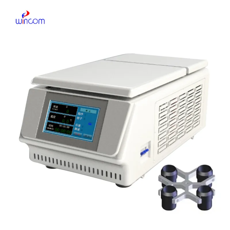
The centrifuge operates quietly and efficiently. It’s compact but surprisingly powerful, making it perfect for daily lab use.
To protect the privacy of our buyers, only public service email domains like Gmail, Yahoo, and MSN will be displayed. Additionally, only a limited portion of the inquiry content will be shown.
We are planning to upgrade our imaging department and would like more information on your mri machin...
We’re interested in your delivery bed for our maternity department. Please send detailed specifica...
E-mail: [email protected]
Tel: +86-731-84176622
+86-731-84136655
Address: Rm.1507,Xinsancheng Plaza. No.58, Renmin Road(E),Changsha,Hunan,China

 af
af
 es
es
 ar
ar
 tr
tr
 sw
sw
 pt
pt
 th
th
 ur
ur
 bn
bn
 ne
ne
 vi
vi
 km
km
 lo
lo
 de
de
 ru
ru
 fi
fi
 nl
nl
 fa
fa
 fr
fr
 ko
ko