The ultrasound gel for fetal doppler has been designed considering the needs of modern health care, providing uninterrupted performance with its rapid image acquisition and high-definition visualization. The robust outer casing and sophisticated temperature control system will make sure that the device continues to work and be trustworthy. Also, the ultrasound gel for fetal doppler is a device that aids the long-term data archiving process for efficient medical record management.

The vast clinical applications of the ultrasound gel for fetal doppler technology made it possible for nephrology to monitor kidney function efficiently and detect abnormalities in kidney structure. In the frontiers of endocrinology, the obtained data can reveal even the smallest nodules in the glands. The ultrasound gel for fetal doppler is also a surgical device for blood flow patterns and vessel integrity.

Through continued innovations in digital technology, the ultrasound gel for fetal doppler can be expected to improve and extend its applications within preventive medicine and telemedicine. The next generation of such technologies will facilitate collaboration among experts in real-time using cloud-imaging solutions. The ultrasound gel for fetal doppler can also work within wearables that include biosensors.

The ultrasound gel for fetal doppler needs to be maintained based on the manufacturer's recommendations. The transducers should be kept in specialized holders. The cleaning agent should be non-corrosive. The electrical contacts should remain dry. Functional tests should be carried out on a regular basis to ensure that the ultrasound gel for fetal doppler functions properly and remains a safe instrument.
With the advanced imaging technology, the ultrasound gel for fetal doppler provides physicians unobstructed and precise images of internal organs. It is employed in the early disease diagnosis as well as in patient tracking. The ultrasound gel for fetal doppler functions by utilizing sound wave reflections to generate dynamic images, qualifying it as an essential tool in modern medical diagnostics. Through the ultrasound gel for fetal doppler, fast, non-invasive testing is facilitated for real-time assessment to support clinical decisions.
Q: What is the primary function of an ultrasound scannert? A: Ultrasound scanners are designed to create real-time images of internal organs, tissues, and blood flow using high-frequency sound waves. Q: How does the ultrasound scannert ensure clear imaging results? A:It uses advanced converter technology and digital processing to enhance image clarity and contrast. Q: In what medical fields is the ultrasound scannert commonly used? A: It is widely used in obstetrics, cardiology, urology, radiology, and emergency medicine. Q: Is the ultrasound scannert safe for repeated use? A: Yes, it is non-invasive and does not emit radiation, making it safe for frequent diagnostic applications. Q: Can the ultrasound scannert store and share imaging data? A: Yes, it supports data storage, retrieval, and digital transfer for easy integration with hospital systems.
This x-ray machine is reliable and easy to operate. Our technicians appreciate how quickly it processes scans, saving valuable time during busy patient hours.
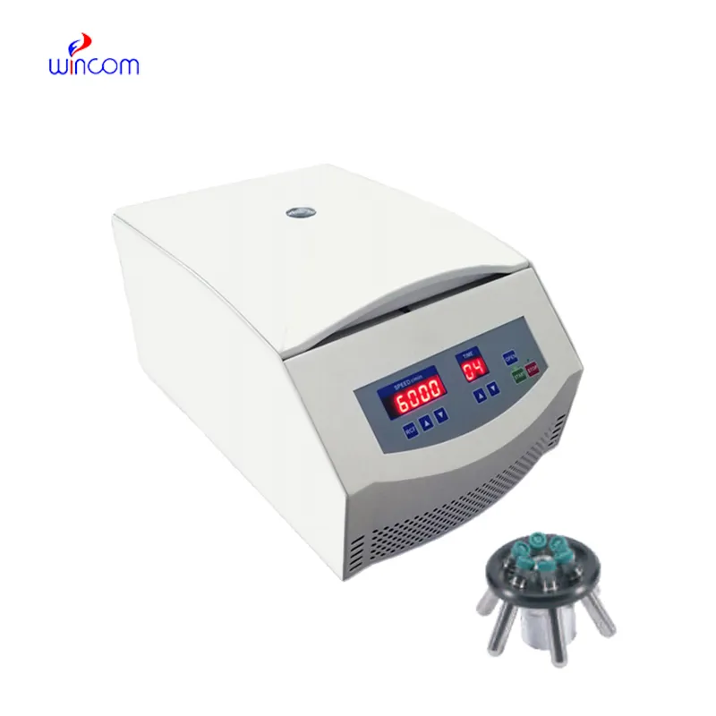
We’ve used this centrifuge for several months now, and it has performed consistently well. The speed control and balance are excellent.
To protect the privacy of our buyers, only public service email domains like Gmail, Yahoo, and MSN will be displayed. Additionally, only a limited portion of the inquiry content will be shown.
I’d like to inquire about your x-ray machine models. Could you provide the technical datasheet, wa...
Could you share the specifications and price for your hospital bed models? We’re looking for adjus...
E-mail: [email protected]
Tel: +86-731-84176622
+86-731-84136655
Address: Rm.1507,Xinsancheng Plaza. No.58, Renmin Road(E),Changsha,Hunan,China

 af
af
 es
es
 ar
ar
 tr
tr
 sw
sw
 pt
pt
 th
th
 ur
ur
 bn
bn
 ne
ne
 vi
vi
 km
km
 lo
lo
 de
de
 ru
ru
 fi
fi
 nl
nl
 fa
fa
 fr
fr
 ko
ko