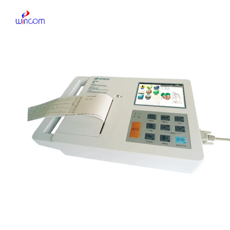
The x ray scanner application comes equipped with an intelligent imaging system that increases grayscale depth and detail understanding. The complex algorithms of the x ray scanner application improve the viewing of subtle lesions and tissues. The x ray scanner application has been designed for high throughput capabilities that promote rapid viewing cycles and convenient data accessibility.

In operating rooms, the x ray scanner application offers real-time imaging assistance in orthopedic and spinal surgeries. It helps surgeons confirm instrument positioning and bone alignment during surgery. The x ray scanner application guarantees accuracy and dependability in real-time intraoperative decision-making.

Future versions of the x ray scanner application will combine energy-efficient technology with high-resolution imaging. Predictive analytics integration will enable early disease detection and personalized screening. Global telemedicine networks will also be enabled by the x ray scanner application, extending access to diagnosis in underserved populations.
Care and maintenance of the x ray scanner application are required to ensure repeat imaging quality and ruggedness. Cable, detector, and collimator faults are averted by periodic checks. The x ray scanner application need to be kept in a dust-free environment with low temperatures to avoid overheating and dust depositing on them. Routine calibration and radiation output monitor checks ensure accurate diagnostic data.
The x ray scanner application has been used heavily in various medical fields due to its ability to offer rapid and precise medical images. The x ray scanner application offers precise images of the various body parts that help in the diagnoses of various conditions such as bone injuries, cancer, and infections. The x ray scanner application uses advanced imaging softwares that offer high contrast images.
Q: What makes an x-ray machine different from a CT scanner? A: An x-ray machine captures a single 2D image, while a CT scanner takes multiple x-rays from different angles to create 3D cross-sectional views. Q: How is image quality measured in an x-ray machine? A: Image quality depends on factors like contrast, resolution, and exposure settings, which are adjusted based on the target area being examined. Q: What power supply does an x-ray machine require? A: Most x-ray machines operate on high-voltage power systems, typically between 40 to 150 kilovolts, depending on their intended use. Q: Can x-ray machines be used for dental imaging? A: Yes, specialized dental x-ray machines provide detailed images of teeth, jaws, and surrounding structures to support oral health assessments. Q: How does digital imaging improve x-ray efficiency? A: Digital systems allow instant image preview, faster diagnosis, and reduced need for retakes, improving workflow efficiency in clinical environments.
I’ve used several microscopes before, but this one stands out for its sturdy design and smooth magnification control.
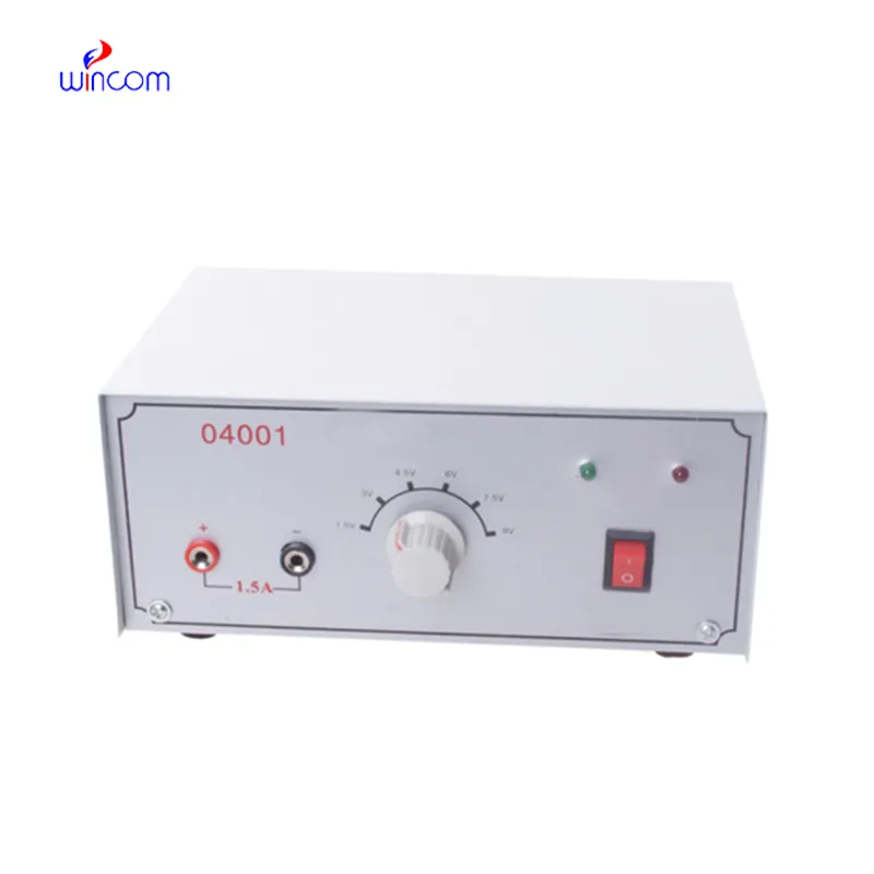
This x-ray machine is reliable and easy to operate. Our technicians appreciate how quickly it processes scans, saving valuable time during busy patient hours.
To protect the privacy of our buyers, only public service email domains like Gmail, Yahoo, and MSN will be displayed. Additionally, only a limited portion of the inquiry content will be shown.
Could you share the specifications and price for your hospital bed models? We’re looking for adjus...
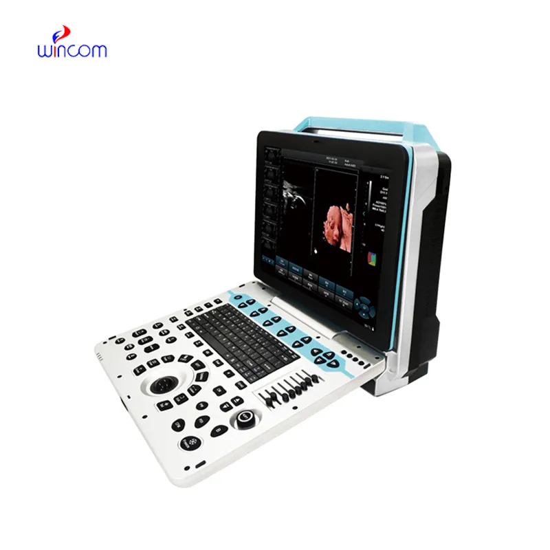
We’re currently sourcing an ultrasound scanner for hospital use. Please send product specification...
E-mail: [email protected]
Tel: +86-731-84176622
+86-731-84136655
Address: Rm.1507,Xinsancheng Plaza. No.58, Renmin Road(E),Changsha,Hunan,China

 af
af
 es
es
 ar
ar
 tr
tr
 sw
sw
 pt
pt
 th
th
 ur
ur
 bn
bn
 ne
ne
 vi
vi
 km
km
 lo
lo
 de
de
 ru
ru
 fi
fi
 nl
nl
 fa
fa
 fr
fr
 ko
ko